
Postintervention monitoring of peripheral arterial disease wound healing using dynamic vascular optical spectroscopy
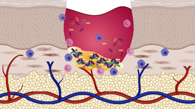
Peripheral arterial disease (PAD) is a vascular disease that is caused by clogging of the arteries due to plaque. The lower legs and feet are often impacted, and symptoms include pain, numbness, difficulty walking, and non-healing wounds.
For many patients, wounds continue not to heal even after they have undergone a surgical revascularization procedure designed to unclog the affected arteries, necessitating another intervention. Determining whether wounds will heal must be done as soon as possible after the first intervention to reduce the duration of a patient’s pain and increase the likelihood of a good outcome.
Currently, the most common methods for monitoring PAD progression (and related wound healing) are the ankle-brachial index (ABI) and ultrasound imaging. The ABI uses the ratio of systolic blood pressure measurements from arteries in the lower extremities to the systolic blood pressure measurement from the brachial artery in the arm. PAD patients generally have pressure ratios that are below a certain threshold (often 0.9). The ABI has low accuracy when monitoring vasculature in diabetic patients and ultrasound imaging has low accuracy when monitoring smaller arteries such as those in the feet. Unfortunately, PAD is often comorbid with diabetes and the affected arteries are commonly in the feet.
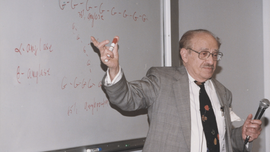
To address the existing limitations of the current technology, a team from NYU Tandon School of Engineering’s department of Biomedical Engineering, including lead author Nisha Maheshwari from Andreas Hielscher's Clinical Biophotonics Laboratory, developed optical imaging technology to assist physicians with monitoring the healing of lower limb ulcers after a surgical intervention.
Dynamic vascular optical spectroscopy (DVOS) is an optical imaging technology that uses light in the red and near-infrared ranges to determine characteristics of blood flow through arteries. In the paper “Postintervention monitoring of peripheral arterial disease wound healing using dynamic vascular optical spectroscopy,” published in the Journal of Biomedical Optics, the team used their DVOS system to monitor a set of 14 patients with PAD that underwent a surgical revascularization procedure. Of these patients, five needed a second intervention due to the persistence of non-healing wounds.
The team was able to correctly categorize the long-term healing and non-healing of wounds in 93% of this patient population within only one month after each patient’s initial intervention. The method outperformed the gold standard methods of ultrasound and ABI. These findings suggest that the DVOS may be able to assist physicians in improving patient outcomes and reducing long-term pain by determining wound outcome earlier than existing technology can.
The authors would like to thank the patients who volunteered for this study for their time and participation. This work was supported in part by the National Heart, Lung, and Blood Institute (Grant No. NHLBI-1R01-HL115336); Wallace H. Coulter Foundation; Society of Vascular Surgery; Columbia University Fu Foundation School of Engineering and Applied Science; and New York University Tandon School of Engineering.
"Maheshwari N, Marone A, Altoé M, Kim SHK, Bajakian DR, Hielscher AH. Postintervention monitoring of peripheral arterial disease wound healing using dynamic vascular optical spectroscopy. J Biomed Opt. 2022 Dec;27(12):125002. doi: 10.1117/1.JBO.27.12.125002. Epub 2022 Dec 24. PMID: 36582192; PMCID: PMC9789744."

Nisha Maheshwari